Non-invasive technique to record involuntary nervous system
The technology could help shed light on the involuntary nervous system’s role in sepsis, PTSD and other inflammatory condition
From the University of San Diego
A research team led by UC San Diego has, for the first time, shown that a wearable, non-invasive device can measure activity in human cervical nerves in clinical settings.
The device records what the team calls Autonomic Neurography (ANG), neural activity from the human vagus and carotid sinus nerves as well as other autonomic nerves found in the skin and muscle of the neck. The vagus nerve is a “superhighway” of the involuntary nervous system, with tendrils extending from the base of the skull through the torso and abdomen to influence digestion, heart rate and the immune system.
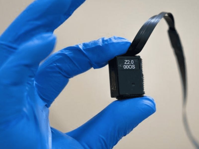
To offer medical professionals a real-time, clinically proven tool for detecting levels of activity in the involuntary nervous system, an early warning sign of a body under stress, the researchers designed a flexible, adhesive-integrated electrode array (as reported in a 2022 Scientific Reports paper). The current study, published July 29, 2024 in Nature Communications Biology, used this approach with the aim of detecting deep neural activity in a simulated clinical hyperinflammatory model.
To replace surgically implanted microelectrodes to monitor or activate the vagus nerve, the new device leverages a powerful technique called “magnetoneurography” to more accurately detect cervical nerve firing non-invasively in real-time. The device detects the magnetic fields arising from activity in the vagus and carotid sinus nerves, which “pulse” to warn the involuntary nervous system of a threat.
Researchers tested the device in nine adult human subjects. Patients had their blood drawn and their plasma tested for baseline levels of inflammation-triggering proteins called cytokines. Next, they were injected with bacteria-sourced toxins called lipopolysaccharides, inducing a temporary hyperinflammatory state in the body that mimicked the inflammation associated with a blood infection.
Inside a magnetically shielded room at the UC San Diego Qualcomm Institute Magnetoencephalography Center, researchers placed sensors from their device at points of the vagus nerve beneath the right ear and over the right carotid artery, where both the vagus nerve and carotid sinus nerve are found. The device monitored heart rate and the magnetic fields arising from nerve activity.
Within half an hour of the patients’ injection with lipopolysaccharides, the device detected changes in their nerve activity below the right ear. Researchers confirmed the rise in nerve activity and the release of inflammatory proteins through blood samples. They also recorded changes in heart rate throughout, as well as a noticeable link between nerve firing at both sites and changes in a particular inflammatory cytokine called tumor necrosis alpha, or TNF-α and the anti-inflammatory cytokine called IL-10.
 If you enjoyed this article, you will like the following ones: don't miss them by subscribing to :